Anatomy Muscles Pelvis / Mri Pelvis Anatomy Free Male Pelvis Axial Anatomy. This mri male pelvis axial cross sectional anatomy tool is absolutely free to use. The anterior compartment includes pectineus, iliopsoas, psoas minor, iliacus. These muscles, including the gluteus maximus and the hamstrings, extend the thigh at the hip in support of the body's weight and propulsion. The pubococcygeus (pc) muscle is the muscle that runs the show in pelvic floor health. The pelvis's frame is made up of the bones of the pelvis, which connect the axial skeleton to the femurs, and therefore acts in weight bearing of the upper body.
A proper kegel exercise means a full contraction and relaxation of the pc muscle. They form a large sheet of skeletal muscle that is thicker in some areas than in others. Rectus femoris muscle, one of the quadriceps muscles on the front of your thigh. Study human anatomy with reliable 3d models & detailed articles. There are many muscles that form the pelvic floor, including puborectalis, pubococcygeus, iliococcygeus and coccygeus.
Budget Pelvis Model With Organs And Pelvic Floor Muscles Anatomystuff Co Uk from www.anatomystuff.co.uk The adductor muscle group, also known as the groin muscles, is a group located on the medial side of the thigh. The main function of the pelvic floor muscles are: Large ligaments, tendons, and muscles around the hip joint hold the bones (ball and socket) in place and keep it from dislocating. The ilium, ischium and the pubic bone. Psoas consists of a pair of deep muscles (psoas major and iliacus) located on each side of the pelvis in the abdomen. The pelvic brim involves the first sacral segment, the iliac and pubis portion, but not the ischium. The pelvic girdle, also known as the hip bone, is composed of three fused bones: Ligaments, tendons, and muscles play an important role in the function of the hip.
The floor of the pelvis is made up of the muscles of the pelvis, which support its contents and maintain urinary and faecal continence.
These muscles are particularly responsible for the support of pelvic organs and maintenance of continence. (2) the levator ani and the coccygeus, which together form the pelvic diaphragm and are associated with the pelvic viscera. See more ideas about anatomy, thoracic, basic image. The levator ani muscles consist of three. The muscles of the femoral region of the lower limb are divided into three compartments. The floor of the pelvis is formed by the two muscles named levator ani and coccygeus. Explore every muscle, bone and organ in 3d However, often they will do more than one movement, assisting another muscle. This mri male pelvis axial cross sectional anatomy tool is absolutely free to use. Attached to the pelvis are muscles of the buttocks, the lower back, and the thighs. Psoas consists of a pair of deep muscles (psoas major and iliacus) located on each side of the pelvis in the abdomen. Ligaments, tendons, and muscles play an important role in the function of the hip. The thigh bone or femur and the pelvis join to form the hip joint.
Use the mouse scroll wheel to move the images up and down alternatively use the tiny arrows (>>) on both side of the image to move the images.>>) on both side of the image to move the images. Attached to the pelvis are muscles of the buttocks, the lower back, and the thighs. The pelvic girdle, also known as the hip bone, is composed of three fused bones: The floor of the pelvis is made up of the muscles of the pelvis, which support its contents and maintain urinary and faecal continence. These muscles, including the gluteus maximus and the hamstrings, extend the thigh at the hip in support of the body's weight and propulsion.
Pelvis Muscle Bone Anatomy 3d Cgtrader from img2.cgtrader.com Piriformis the piriformis is a triangular muscle 1 on either side on the very front of the posterior wall of true pelvis. These muscles originate near the anteroinferior external surface of the bony pelvis and insert at the linea aspera. It attaches inferiorly (underneath/below) to the long thick strip of fascia, known as. There are many muscles that form the pelvic floor, including puborectalis, pubococcygeus, iliococcygeus and coccygeus. The thigh bone or femur and the pelvis join to form the hip joint. These muscles origin in continuity from the body of the pubis, along a tendinous arch over the obturator internus fascia, and the ischial spine. Psoas consists of a pair of deep muscles (psoas major and iliacus) located on each side of the pelvis in the abdomen. The function of the pelvic floor is to help assist with child birth, prevent incontinence and support organs within the pelvis.
These muscles are particularly responsible for the support of pelvic organs and maintenance of continence.
(1) the obturator internus and the the fascia of the obturator internus covers the pelvic surface of, and is attached around. The pelvic girdle and pelvic spine. To support the abdominal and pelvic viscera The anterior or extensor, medial or adductor, and posterior or flexor compartments. The pelvic floor muscles provide foundational support for the intestines and bladder. Each compartment is separated from the others by an intermuscular septum that runs from the fascia lata to the linea aspera of the femur. The muscles of the pelvic floor are collectively referred to as the levator ani and coccygeus muscles. It can be described as one of the bodies diaphragms. Ligaments, tendons, and muscles play an important role in the function of the hip. The tensor fasciae latae (tfl) is a small muscle on the outside of the hip. The deepest layer is the pelvic diaphragm, the muscles that make up the pelvic diaphragm are pubococcygeus, puborectalis, pubourethralis, iliococcygeus and ischiococcygeus. The pelvis's frame is made up of the bones of the pelvis, which connect the axial skeleton to the femurs, and therefore acts in weight bearing of the upper body. The pelvic inlet involves three of the four units of which the bone pelvis is composed.
The levator ani muscles consist of three. The pubococcygeus (pc) muscle is the muscle that runs the show in pelvic floor health. Explore every muscle, bone and organ in 3d Piriformis the piriformis is a triangular muscle 1 on either side on the very front of the posterior wall of true pelvis. It attaches inferiorly (underneath/below) to the long thick strip of fascia, known as.
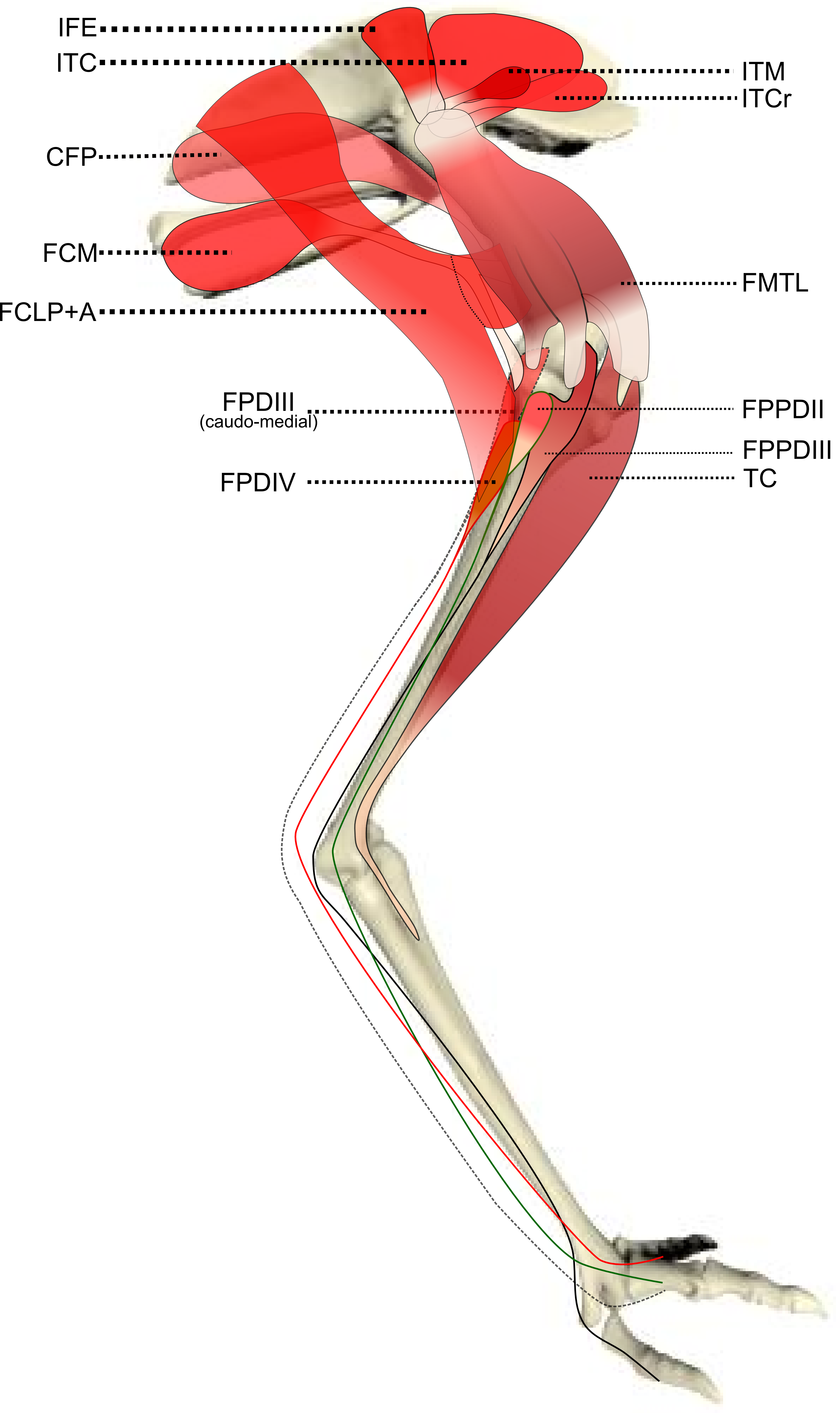
Ontogenetic Scaling Patterns And Functional Anatomy Of The Pelvic Limb Musculature In Emus Dromaius Novaehollandiae Peerj from dfzljdn9uc3pi.cloudfront.net The joint's natural development and mechanical loading pattern change with age to accommodate the growing physiological stresses and impacts on the pelvic and hip regions. Helps improve kegel and pelvic floor. The muscles of the pelvic floor are collectively referred to as the levator ani and coccygeus muscles. To support the abdominal and pelvic viscera Muscles of the male & female pelvic floor | anatomy model These muscles arise from the hip, spine, and proximal femur. This mri male pelvis axial cross sectional anatomy tool is absolutely free to use. Pelvic floor anatomy that is easy to understand!
(1) the obturator internus and the piriformis, which are muscles of the lower extremity, and will be described with these (pages 476 and 477);
Muscles of the male & female pelvic floor | anatomy model Choose from 500 different sets of flashcards about anatomy muscles pelvis on quizlet. The anterior compartment includes pectineus, iliopsoas, psoas minor, iliacus. The pelvic floor muscles provide foundational support for the intestines and bladder. Large ligaments, tendons, and muscles around the hip joint hold the bones (ball and socket) in place and keep it from dislocating. The ilium, ischium and the pubic bone. The deepest layer is the pelvic diaphragm, the muscles that make up the pelvic diaphragm are pubococcygeus, puborectalis, pubourethralis, iliococcygeus and ischiococcygeus. (1) the obturator internus and the piriformis, which are muscles of the lower extremity, and will be described with these (pages 476 and 477); The pelvis's frame is made up of the bones of the pelvis, which connect the axial skeleton to the femurs, and therefore acts in weight bearing of the upper body. The joint's natural development and mechanical loading pattern change with age to accommodate the growing physiological stresses and impacts on the pelvic and hip regions. The anterior or extensor, medial or adductor, and posterior or flexor compartments. The pelvic brim involves the first sacral segment, the iliac and pubis portion, but not the ischium. The muscles of the pelvic floor are collectively referred to as the levator ani and coccygeus muscles.